Research Images
SERCEB investigators who would like to submit images of their research for display on these pages are welcome and encouraged to do so. Please e-mail jpeg files to Elaine Fitzsimons.
 |
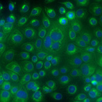
Yellow Fever Virus
Contributed by Mariano Garcia-Blanco
|
 |
Cowpox
Cowpox virus infected chorioallantoic membranes showing a mutant “white” pock amidst wild type virus pocks. The mutant virus is deleted in a gene which controls inflammation. The “white” is due to inflammatory heterophi (neutrophil) influx which is absent in wild type pocks.
Contributed by Richard Moyer
|

|
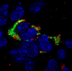
Francisella tularensis
Francisella tularensis (green) infected lung epithelial cell labeled with anti-proSPB (red) and DAPI (blue) stained nuclei.
Contributed by Tom Kawula |
 |
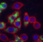
Francisella tularensis
Francisella tularensis (green) infected macrophages 24 hours post inoculation.
Contributed by Tom Kawula |